| LOWER LIMB BLOCKS WITH NERVE STIMULATOR |
| Femoral Nerve Block Three in one Nerve Block Lateral Femoral Cutaneous Nerve |
| Obturator Nerve Popleteal Block Classic approach for sciatic nerve block |
| Sciatic Nerve Block Anterior approach Ankle Block Saphenous Nerve Block |
 |
Dr.R.Silamban(Asst. Professor) |
| FEMORAL NERVE BLOCK | ||||||||||
| Position:Supine | ||||||||||
| Site of Needle Entry:A point 2 cms below the inguinal ligament and 1cm lateral to the femoral artery. | ||||||||||
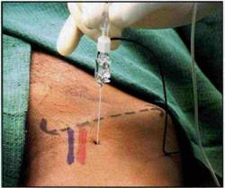 | ||||||||||
| Technique: A 4cm needle is entered at the above point perpendicular to the skin to pierce the iliopectineal fascia and fascia lata and parasthesia is elicited. If nerve locator is used the motor response depends on division of the femoral nerve that is stimulated. If the anterior division is stimulated the vasti respond. It is always better to have a motor response of the posterior division for a complete block especially if surgery is around the knee joint. | ||||||||||
| ||||||||||
| Volume: 20ml | ||||||||||
| Pearls: | ||||||||||
| ||||||||||
| THREE IN ONE NERVE BLOCK (INGUINAL PERIVASCULAR BLOCK) | ||||||||||||||||
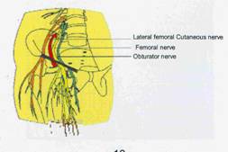
| It is based on the concept that injection of local anaesthetic near the femoral nerve in higher volumes causes the local anaesthetic to track proximally along the fascial planes between the iliacus and psoas muscles to reach the lumbar plexus roots. | ||||||||||||||||
| This blocks all the three major nerves arising from the lumbarplexus namely the lateral cutaneous nerve of thigh, the femoral nerve and the obturator nerve. | ||||||||||||||||
 | ||||||||||||||||
| Position: Supine | ||||||||||||||||
| Site of Needle Entry: Similar to femoral nerve block but the direction of needle entry may be about 45 degree to 60 degree to the skin especially when a continous catheter is planned. The angulation of the needle facilitates catheter insertion. | ||||||||||||||||
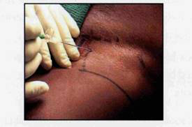 | ||||||||||||||||
| Technique: Once the femoral nerve is located with the nerve locator the local anaesthetic is given while giving distal pressure. The drug may also be milked cephalad to facilitate travel along the iliopsoas muscle plane. Alternatively a catheter may be introeduced through a suitable needle to lie within the iliopsoas muscle plane near the lumbar nerve roots as shown in the figure. Catheter is inserted for 10-15cms. The muscle plane may be expanded with local anesthetic or normal saline prior to cathether insertion. | ||||||||||||||||
| Volume:30-40ml | ||||||||||||||||
| Pearls | ||||||||||||||||
| ||||||||||||||||
| LATERAL CUTANEOUS NERVE OF THIGH | |||||||
| Root Value: L2, L3 | |||||||
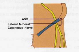
| Course:Takes a very lateral course in the pelvis to come behind the lateral end of the inguinal ligament and lie beneath the fascia lata. Within 2.5cm of entering the thigh it divides into multiple small branches to supply the skin on the lateral side of thigh. | |||||||
 | |||||||
| Position: Supine | |||||||
| Site of Needle Entry:2cm medial and inferior to the anterior superior iliac spine | |||||||
| Technique: At the above point a 4cm needle is advanced perpendicular to the skin till a “pop off” is felt once the fascia lata is pierced. 8ml is given here. The needle is withdrawn above the fasica lata and additional 3 ml of drug is given here to block some cutaneous branches. | |||||||
| Volume:10 to 12 ml | |||||||
| Pearls | |||||||
| |||||||
| OBTURATOR NERVE | |||||||
| Root Value: L2, L3 & L4 | |||||||
| Course: Upper part of the nerve lies in the pelvis and enters the thigh by passing through the obturator foramen. Just outside the foramen it divides into anterior and posterior divisions. | |||||||
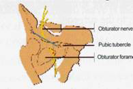 | |||||||
| Course:They supply the adductor muscle of the thigh and also gives a small branch to the capsule of the knee joint. | |||||||
| Position: Supine | |||||||
| Site of Needle Entry: 1.5cm below and lateral to pubic tubercle. | |||||||
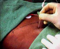 | |||||||
| Technique: : A 10cm needle is advanced perpendicularly till the ramus of pubis is contacted. It is then withdrawn and redirected in a lateral direction to pierce the obturator membrane which is felt as a give way and parasthesia is elicited. If a nerve locator is usedcontractions of the adductors of the thigh is elicited. The nerve is usually located at a depth of 6-7cms from the skin. | |||||||
| Volume:10 ml | |||||||
| Pearls | |||||||
| |||||||
| POPLETEAL BLOCK | ||||||||||
| Involves blocking the sciatic nerve in the popliteal fossa. Useful block in foot and ankle surgery. Combined with saphenous nerve block for full analgesia of the foot and ankle. | ||||||||||
| Position: Prone | ||||||||||
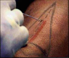
| Site of Needle Entry:A point 5cms above the popliteal skin crease and 1 cm lateral to the line bifurcating the popliteal fossa. | ||||||||||
| Technique: A7.5cm needle is preferred. It is advanced at an angle of 45 degree to 60 degree to the skin in a cephalad direction. At a depth of 3.5 to 5cm parasthesia or motor response is elicited and drug in injected. It is advisable to have a tibial motor response of plantar flexion and foot inversion. | ||||||||||
 | ||||||||||
| Volume:30 ml | ||||||||||
| Pearls | ||||||||||
| ||||||||||
| SCIATIC NERVE BLOCK | |||||||
There are two common approaches | |||||||
| |||||||
| CLASSIC APPROACH FOR SCIATIC NERVE BLOCK | |||||||||||||||||||
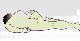
| Position: :Patient is placed laterally. Side to be blocked is nondependent. Dependent leg is extended and nondependent leg is flexed to 90 degree. Heel is placed apposing the knee of the dependent leg. | |||||||||||||||||||
 | |||||||||||||||||||
| Site of Needle Entry: | |||||||||||||||||||
| |||||||||||||||||||
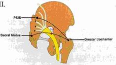 | |||||||||||||||||||
| Patient is placed laterally. Side to be blocked is nondependent. Dependent leg is extended and nondependent leg is flexed to 90 degree. Heel is placed apposing the knee of the dependent leg. | |||||||||||||||||||
| Technique: A12 to 15cm needle is entered from the above point perpendicular to the skin towards an imaginary point where femoral vessels go under the inguinal ligament. If the bone is contacted before parasthesia or motor response withdraw the needle and | |||||||||||||||||||
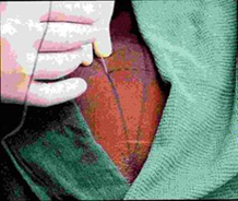 | |||||||||||||||||||
| redirect it along the second line till parasthesia or motor response is obtained. Do not advance the needle for more than 2cm after bony contact. The nerve is usually located at adepth of 8cms in a moderately built patient. Paresthesia is more commonly felt in the peroneal territory. The motor response to be anticipated is dorsiflexion and eversion of foot. | |||||||||||||||||||
| Volume:15-20 ml | |||||||||||||||||||
| Pearls | |||||||||||||||||||
| |||||||||||||||||||
| SCIATIC NERVE BLOCK ANTERIOR APPROACH | ||||||||||
| Position: Supine with leg in a slightly abducted position | ||||||||||
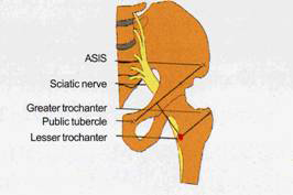 | ||||||||||
| Site of Needle Entry: | ||||||||||
| ||||||||||
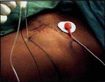
| Technique: A 12 or 15cm needle is inserted perpendicular to the skin. At about 5 to 6 cm it contacts the femur. The needle is withdrawn and directed medially and if necessary little cephalad to locate the sciatic nerve. About 4cms past the femur, parasthesia or motor response is elicited. Inversion/eversion of foot or dorsiflexion / plantar flexion of ankle is the motor response obtained, depending on whether the tibial or common peroneal is stimulated. | ||||||||||
 | ||||||||||
| Volume:15 ml | ||||||||||
| Pearls | ||||||||||
| ||||||||||
| ANKLE BLOCK | ||||||||||||||||
| Involves blocking the following five nerves around ankle joint | ||||||||||||||||
| ||||||||||||||||
| Technique: | ||||||||||||||||
 | ||||||||||||||||
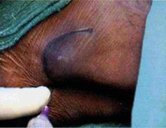
| Medial:The posterior tibial artery is palpated near the medial malleolus. Needle is inserted close to the artery to elicit parasthesia. If it is difficult to elicit paresthesia, advance the needle further to hitch the medial malleolus. Then withdraw by 1cm and give 3-4ml of LA. | ||||||||||||||||
 | ||||||||||||||||
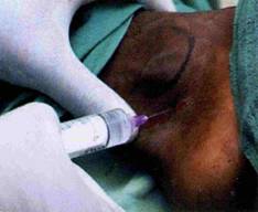
| Lateral:The needle is inserted just below the lateral malleolus to elicit paresthesia. If the paresthesia cannot be elicited, hitch the medial malleolus and withdrfaw by 1.5cm and give 3-4ml of LA. | ||||||||||||||||
 | ||||||||||||||||
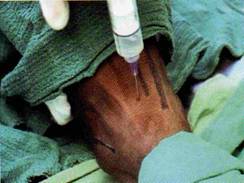
| Dorsal:Insert needle between the anterior tibial artery and Extensor Halucis Longus tendon on the line jointing the two malleolus. Once flexor retinaculum is pierced inject 3 to 4 of local anaesthetic to block the deep peroneal nerve. From this point a subcutaneous weal is raised aznd drug is given in a medial and lateral direction to block the saphenous and superficial peroneal nerve. | ||||||||||||||||
 | ||||||||||||||||
| Pearls | ||||||||||||||||
| ||||||||||||||||
| SAPHENOUS NERVE BLOCK | ||||
| Not used in isolation. Very often combined with sciatic nerve block for complete analgesia of ankle and foot. It is a branch of the posterior division of the femoral nerve. | ||||
| Anatomy | ||||
| It becomes superficial by piercing the deep fascia between the gracilis and sartorius muscle to run in front of the great saphenous vein. | ||||
| Position: Patient supine and knee bent to 45 degree | ||||
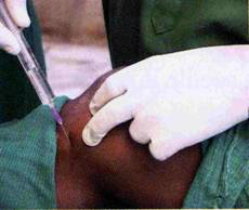 | ||||
| Technique: A sausage of local anaesthetic is injected jujst distal to the medial surface of the tibial tuberosity in the subcutaneous plane. | ||||
| Volume: 5-6ml | ||||
| Pearls | ||||
| ||||
| Download Hand Book On | ||||
| Lower Limb Blocks Nerve Blocks [ Click Here ] | ||||