Conference Lectures
Increased end-tidal carbondioxide - a diagnostic dilemma.
Dr. Indira Gurajala, Associate Professor
Department of Anaesthesiology and Critical Care
Nizams Institute of Medical Sciences, Hyderabad
ASA guidelines recommend that continual end-tidal CO2 analysis should be performed until the endotracheal tube or LMA device is removed or the patient is transferred to a postoperative care location. The simplest use of measured ETCO2 is to follow the changes in PaCO2 level. Usually the difference between the arterial and ETCO2 is 4-6 mmHg. Anaesthesiologists commonly use a capnogram to confirm tracheal intubation and to exclude esophageal intubation. However, the end tidal carbondioxide (ETCO2) levelshown by the capnogram and the pattern of capnogram can also indicate many hemodynamic and respiratory conditions.Also conditions leading to an increased arterial - end – tidal CO2 should be recognized and treated.
NORMAL CAPNOGRAPH :Capnography is the graphic display of instantaneous CO2 partial pressure versus time during the respiratory cycle. The waveform should be examined systematically for height, frequency, rhythm, baseline, and shape. Height depends on the end-tidal CO2. Frequency depends on the respiratory rate.
The baseline is normally zero. An elevated baseline can result from deliberate administration of CO2, rebreathing, exhausted absorbent, a contaminated sample cell, or an incompetent expiratory unidirectional valve. The baseline may or may not be elevated with an incompetent inspiratory unidirectional valve.
The shape of the normal waveform: Only one shape (top hat or sine wave) is considered normal.( Figure .1)
Phase I (inspiratory baseline) begins at E and is normally zero, reflecting inspired gas, which is normally devoid of CO2.
Phase II (expiratory upstroke) begins at B and continues to C. This rapid S-shaped upswing represents the transition from gas from the dead space that does not participate in gas exchange and alveolar gas that contains CO2.
Phase III begins at C and continues to just before D. As gas coming almost entirely from alveoli is exhaled, a plateau is normally seen. If a plateau is not seen, the maximum value obtained may not be equivalent to the end-tidal level and the correlation between arterial and end-tidal CO2 is not likely to be good. The slope of this phase is increased by ventilation perfusion abnormalities in the lung as well as external factors such as a kinked tracheal tube. The very last portion of Phase III, identified by D, is referred to as the end-tidal point. The CO2 level here is normally at its maximum. In normal individuals, this is 5% to 5.5%, or 35 to 40 torr. The angle between Phases II and III is called the α (takeoff, elevation) angle. Normally, it is between 100 and 110 degrees. It is decreased with obstructive lung disease, as the dead space volume takes longer to be exhaled. The slope of Phase III depends on the lung's ventilation-perfusion status. Airway obstruction and PEEP cause an increased slope and a larger α angle. Other factors that affect the angle are the capnometer's response time, sweep speed, and the respiratory cycle time. The angle between the end of Phase III and the descending limb of the capnogram is called the β angle. Normally, it is approximately 90 degrees. The angle will be increased with rebreathing and with prolonged response time compared with the respiratory cycle time, particularly in children. The angle will be decreased if the slope of Phase III is increased. In Phase IV, the patient inhales. Normally, CO2 falls abruptly to zero and remains at zero until the next exhalation.

Figure .1.Normal carbon dioxide waveform.
Carbon dioxide is produced in the body tissues, conveyed by the circulatory system to the lungs, excreted by the lungs, and removed by the breathing system. Therefore, changes in respired CO2(either increase or decrease) may reflect alterations in metabolism, circulation, respiration, or the breathing system. There are several causes for increase in EtCO2 which are as follows.
- Metabolism
Monitoring CO2 elimination gives an indication of the patient's metabolic rate. A change in end-tidal CO2 is a reliable indicator of metabolism only in mechanically ventilated subjects. For spontaneously breathing patients, PetCO2 may not increase with increased metabolism because of compensatory hyperventilation by the patient. Causes of increased ETCO2 due to increase include
- Increased temperature
- Thyroid storm
- Shivering
- Convulsions
- Excessive catecholamine production or administration
- Blood or bicarbonate administration
- Release of an arterial clamp or tourniquet
- Parenteral hyperalimentation.
- CO2 absorption from CO2used to inflate the peritoneal cavity during laparoscopy, the pleural cavity during thoracoscopy, a joint during arthroscopy or to increase visualization for endoscopic vein harvest.
- Malignant hyperthermia: The increase in ETCO2 occurs early, before the rise in temperature. Early detection of this syndrome is one of the most important reasons for routinely monitoring CO2. Capnometry can be used to monitor the effectiveness of treatment.
The increase in ETCO2 due to hypermetabolism is usually not associated with changes in the arterial to end tidal gradient or increased inspiratory CO2 or airway pressures and the waveform is normal on capnograph.
- Circulation
End-tidal CO2 increases with increased cardiac output if ventilation remains constant.
- During resuscitation, exhaled CO2 is a better guide to the effectiveness of resuscitation measures than the electrocardiogram (ECG), pulse, or blood pressure.However, if high-dose epinephrine or bicarbonate is used, end-tidal CO2 is not a good resuscitation indicator. End-tidal CO2 levels may be of use in predicting the outcome of resuscitation and the resolution of a pulmonary embolus.
- In patients undergoing Blalock Taussig shunt, flow across the shunt causes rapid increase in ETCO2 as more blood is delivered to the lungs.
- Respiration
Carbon dioxide monitoring gives information about the rate, frequency, and depth of respiration. Some respiratory causes of increased end-tidal CO2 are.
- Rebreathing may be detected by a rising inspired CO2 level, elevated baseline in the waveform and normal end tidal to alveolar gradient .
- Accidental endobronchial intubation may result in a transient fall or rise in end-tidal CO2.
- Upper airway obstruction is detected by an abnormal waveform and an elevated end tidal to alveolar gradient.
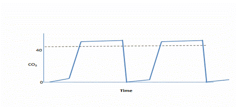
- Hypoventilation has a normal waveform in the capnogram with no inspiratory CO2 and normal end tidal to alveolar gradient (Figure.2).

Figure 2.Elevated end-tidal CO2 with good alveolar plateau may be caused by hypoventilation or increased CO2 delivery to the lungs (due to hypermetabolism or increased cardiac output).
- Equipment Function
A problem with the breathing system can cause an inspired CO2 level greater than zero (Figure .3).
Such problems include
- Leaks
- Faulty or exhausted absorbent
- Channeling
- A bypassed absorber
- Increased dead space

Figure.3.The baseline is elevated, and the waveform is normal in shape. This may be caused by an incompetent expiratory valve or exhausted absorbent in the circle system; insufficient fresh gas flow to a Mapleson system; problems with the inner tube of a Bain system; deliberate addition of CO2 to the fresh gas; or in some cases, an incompetent inspiratory valve.
- Low fresh gas flow to a mapleson system,
- A defect in the inner tube of a bain system,
- Accidental administration of CO2, and
- A defective non rebreathing valve
- Carbon dioxide analysis can be used to detect disconnected oxygen tubing to a mask over the face during local or regional anesthesia. If the oxygen source becomes detached there will be a rise in CO2 because of rebreathing.
- Incompetent unidirectional valves are an inherent danger of the circle system.
- An incompetent expiratory valve allows reverse flow of gas that contains CO2 from the expiratory limb during the inspiratory phase, resulting in an elevated baseline on the capnogram.
- If the inspiratory valve is incompetent, CO2 will enter the inspiratory limb during exhalation. During the next inspiration, CO2 will be rebreathed. This will cause the plateau on the capnogram to be lengthened and a decrease in the steepness of the inspiratory downslope. An increase in the baseline may not be seen. (Figure.4)

Figure.4. Incompetent inspiratory unidirectional valve. The waveform shows a prolonged plateau and a slanting inspiratory downstroke. The inspiratory phase is shortened, and the baseline may or may not reach zero, depending on the fresh gas flow.
Table.1 lists the causes of increased CO2 elimination and ETCO2
|
Waveform on capnograph |
ETCO2 |
Inspired CO2 |
Endtidal to arterial gradient |
1.Capnography and capnometry with increased CO2 production |
||||
a.Absorption of CO2 from peritoneal cavity |
Normal |
⇑ |
0 |
Normal |
b.Injection of NaHCO3 |
Normal |
⇑ |
0 |
Normal |
c.Pain, anxiety, shivering |
Normal |
⇑ |
0 |
Normal |
d.Convulsions |
Normal |
⇑ |
0 |
Normal |
e.Hyperthermia |
Normal |
⇑ |
0 |
Normal |
f.Increased transport of CO2 to the lungs (after release of tourniquet etc) |
Normal |
⇑ |
0 |
Normal |
g. Increased muscle tone |
Normal |
⇑ |
0 |
Normal |
2.Capnography and capnometry as result of circulatory changes |
||||
a.Restoration of circulation after arrest |
Normal |
⇑ |
0 |
Normal |
b.Venous CO2 embolism |
Normal |
⇑ transiently |
0 |
Normal |
3.Capnography and capnometry with respiratory problems |
||||
a.Hypoventilation |
Normal |
⇑ |
0 |
Normal |
b.Upper airway obstruction |
Abnormal |
⇑ |
0 |
Normal |
c.Rebreathing |
Baseline elevated |
⇑ |
⇑ |
⇑ |
4.Capnography and capnometry alterations with equipment |
||||
a.Increased apparatus dead space |
Baseline elevated |
⇑ |
⇑ |
Normal |
b.Rebreathing with circle system |
Baseline elevated |
⇑ |
⇑ |
Normal |
c.Rebreathing with Mapleson system |
Baseline elevated |
⇑ |
⇑ |
Decreased |
d.Rebreathing due to malfunctioning nonrebreathing valve |
Baseline elevated |
⇑ |
⇑ |
Decreased |
e.Obstruction to expiration in the breathing system |
|
|
0 |
Decreased |
TAKE HOME POINTS
- Capnography is a standard of care for every general anaesthetic because it monitors the adequacy of ventilation
- Production of ETCO2 requires both ventilation and perfusion
- Capnography can noninvasively detect many of the catastrophic problems that occur during anesthesia.
- Analysis of the shape of the waveform may give useful information on changes in the patient’s condition, the anesthesia circuit or the effects of surgery .